
Dermoscopy.
Dermoscopy is a hand-held magnification technique that lets your GP see features of the skin invisible to the naked eye. It substantially improves the accuracy of skin cancer detection and is used throughout Clarion skin checks and lesion assessments.
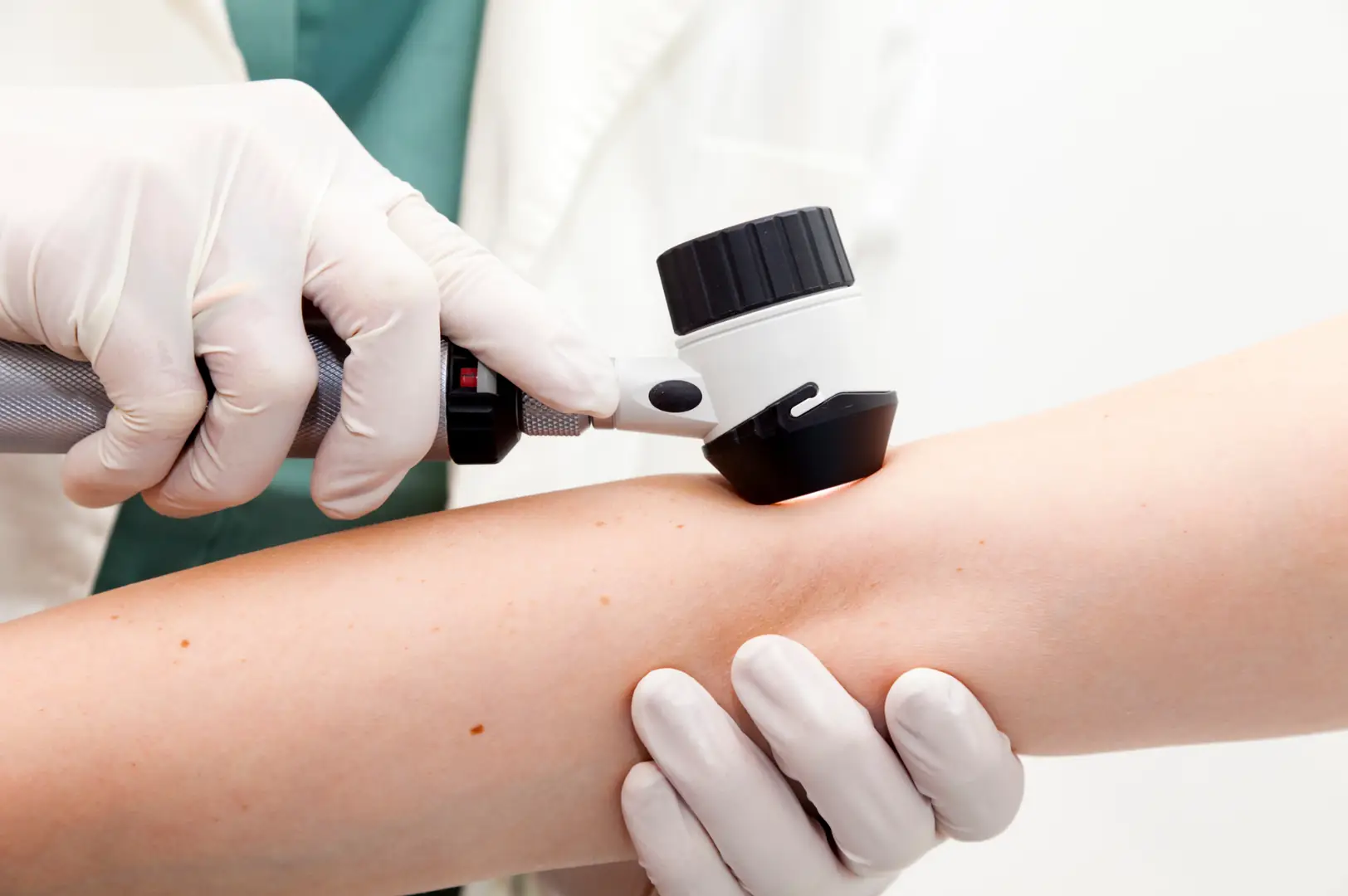
A closer look under the surface.
Dermoscopy uses a hand-held device (a dermatoscope) that combines magnification with polarised light to reveal features of the skin that can't be seen with a standard visual check. The technique lets a trained GP look through the surface of the skin at the structures underneath, which is where the diagnostic clues to skin cancer often live.
Dermoscopy has been shown in multiple studies to substantially improve the accuracy of skin cancer detection compared with visual inspection alone. It reduces both missed cancers and unnecessary biopsies of benign lesions.
At Clarion, dermoscopy is used routinely as part of skin checks and single-lesion assessments. Your GP examines each area of concern under the dermatoscope, looking for specific patterns that indicate whether a lesion is benign, whether it needs monitoring, or whether it needs biopsy or excision.
Four common situations.
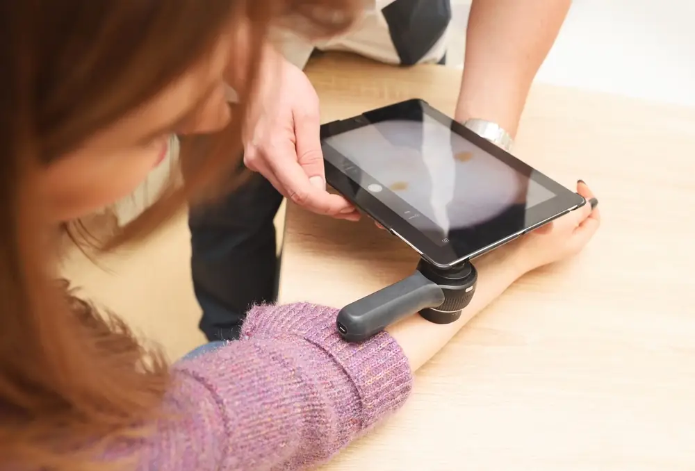
Any spot that needs closer inspection is examined under the dermatoscope. This is standard practice at Clarion.
If you've noticed a specific spot that's new or changing, your GP can examine it dermoscopically as part of a shorter consultation.
Some spots don't warrant excision but need to be watched. Dermoscopy allows precise monitoring for change over time. Digital dermoscopy images can be stored and compared at future checks.
Before excising a lesion, dermoscopy helps confirm the diagnosis and margins.
A decision-support tool.
What dermoscopy can do. Substantially improve the accuracy of skin cancer detection. Identify features that distinguish melanoma from benign moles, and non-melanoma skin cancers from benign lesions. Support decisions about which spots need biopsy and which can be monitored or left alone.
What dermoscopy cannot do. Provide a definitive diagnosis without histology. Any lesion that looks concerning under dermoscopy still needs a biopsy for final confirmation. Dermoscopy is a decision-support tool, not a substitute for pathology.
Non-melanoma cancers. Basal cell carcinoma and squamous cell carcinoma have specific dermoscopic features that support diagnosis, but as with melanoma, histology is required for confirmation.
No prep, no pain.
You don't need to prepare for dermoscopy. It's not painful, requires no injections, and doesn't disrupt your day.
During your appointment, your GP:
- Examines the lesion or lesions visually first.
- Applies the dermatoscope to the skin, sometimes with a thin layer of alcohol or gel to reduce surface reflection.
- Examines features under magnification and polarised light.
- May take a photograph for records or for comparison over time.
- Explains what was seen and what happens next.
If the lesion is clearly benign, no further action is needed. If it needs monitoring, an interval is agreed. If it needs biopsy or excision, the plan is discussed with you.